Article by: Irmazian Abd Shukor (Science Officer, Microscopy Unit)
Electron microscopy is essential in diagnosing renal disease. The Transmission Electron Microscope (TEM), with its ability to visualize structures at the nanometer scale, provides crucial insights into the morphology of renal tissues, thereby enhancing diagnostic accuracy and guiding treatment decisions. Many consider electron microscopy (EM) a necessity for all kidney biopsies; however, EM facilities are limited. At the IBS Microscopy Unit, we offer Transmission Electron Microscope (TEM) services to support the analysis needed for diagnosing renal disease.
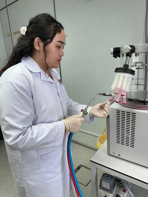
Samples were collected in the hospital using a biopsy technique and sent to the lab in a fixative. The targeted area for pathological diagnosis is the renal glomerulus. The samples will be processed using our lab-developed method and analysed using TEM. Pathologists will collaborate during the analysis to interpret the results.
The high magnification of the Transmission Electron Microscope (TEM) enables impossible observations with light microscopy. More detailed ultrastructure of the renal can be observed such as:
- thickness of the glomerular basement membrane;
- condition of the podocyte food process;
- any complex deposition along the glomerular basement membrane; or
- any other structural abnormalities
Challenges and Limitations
Despite its advantages, EM in renal pathology is not without challenges:
- Technical Complexity: Preparing renal tissue samples for EM requires precise techniques to preserve ultrastructure. This includes fixing, embedding, and sectioning the tissue, which can be technically demanding and time-consuming.
- Interpretation Expertise: EM requires specialized knowledge to interpret the intricate details of cellular structures. Pathologists must be skilled in both the technique and in recognizing patterns associated with various renal diseases.
- Availability: The high cost and complexity of EM equipment limit its availability, particularly in resource-limited settings.
Conclusion
Electron microscopy is a powerful tool in diagnostic renal pathology, offering insights unattainable through conventional light microscopy. By revealing the ultrastructural features of renal tissues, TEM enhances diagnostic precision for a range of glomerular and tubular diseases, ultimately guiding more effective patient management. As technology advances, TEM is anticipated to continue playing a crucial role in diagnosing and understanding renal diseases, bridging gaps that other diagnostic modalities cannot address.
Acknowledgment
The author would like to acknowledge Dr. Fauzah binti Abd Ghani, Renal Pathologist from Hospital Sultan Abdul Aziz Shah (HSAAS), UPM as the expert in renal pathology interpretations.


Date of Input: 29/10/2024 | Updated: 29/10/2024 | azah
MEDIA SHARING