Author: Dr. Kartini Jasni
There are a few techniques available for the assessment of the estrus cycle in animals. The most common method isa visual assessment of the vagina carried out by an experienced personnel member who can differentiate between the estrus stages by examining the appearance of the vagina.
For example, in rats. This method can be done when the animal is approximately 6 to 8 weeks of age. By gently lifting the rat’s hind region, personnel would be able to visualise the vagina and photograph it, if necessary, for record purposes. During proestrus phase, the vagina of a rat opening appears full, swollen and moist. Proestrus is a phase that the estrus phase appears like the proestrus phase, except the vagina has lesser pink in colour, less swollen and less moist. In the metestrus phase, the vagina appears to be pale and dry, while the vagina appears to be very wet during the diestrus phase.
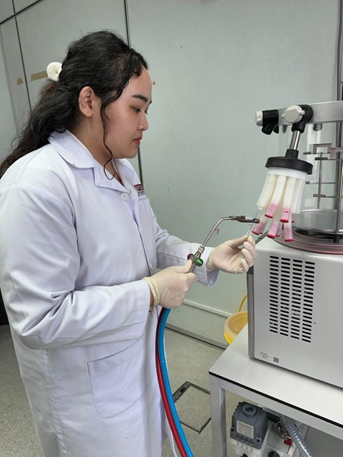
The other method to assess the estrus cycle in animals is the collecting vaginal cells via vaginal lavage to examine vaginal cytology. This method is widely accepted and easy to perform. By using Phosphate Buffer Saline (PBS), disposable pipette, glass slide and microscope, personnel would be able to evaluate a female rat estrus cycle. First, they would restrain a female rat by lifting its hind region, then slowly pumping 0.2mL of the PBS into the vaginal cavity using a pipette. The solution is aspirated, and the process is repeated 4 to 5 times before placing 1 drop of the liquid on the glass slide. The glass slide is covered with a cover slip and visualised under the microscope. Some personnel prefer to wait for the liquid to dry before staining with Papinicolaou stain2. This staining method enhances the accuracy in determination of various stage of estrus cycle in rats which is further useful in studying the effect of test compounds on reproductive system in rats and slides can be keep further for future reference.
Cells from different estrus stages appear differently under the microscope. Cells during the proestrus shows numerous nucleated cells and uniform in size. Cells can be individual or in clusters. Some anucleated cornified epithelial cells and leukocytes can be present as well.
As for estrus, presence of numerous anucleated cornified epithelial cells. Cytoplasms are granular and cells are irregular in shape. No leukocytes but occasionally has bacteria. Example of vaginal smear in estrus phase as attached below.
At metestrus vaginal cytology appears to have numerous leukocytes, few large non granular and anucleated cornified cells.
Diestrus has numerous polymorphonuclear leukocytes and a very few epithelial cells.
In conclusion, understanding all these four stages play important role in reproductive research or for breeding of the animals for research purposes.
Acknowledgement:
The author would like to acknowledge Dr Chau De Ming for his useful suggestions and other COMeT’s staff for all the help during the procedure.
References:
- Staging of the estrous cycle and induction of estrus in experimental rodents: an update. Fertility and Research Practice (2020) Ayodeji Folorunsho Ajayi & Roland Eghoghosoa Akhigbe.
- A Comparative Study on Staining Techniques for Vaginal Exfoliative Cytology of Rat, Journal of Pharmacology and Clinical Research (2017), MR Srivinasan et.al

Date of Input: 20/05/2025 | Updated: 20/05/2025 | azah
MEDIA SHARING